Planned examinations
ExaminationsBelow you will find a detailed insight into all the examinations carried out as part of the COR-PHYS study and their benefits.
Cardiopulmonary exercise testing (Endurance test)
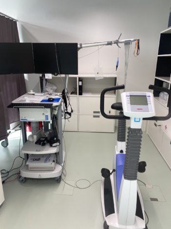
Aim of the examination: Cardiopulmonary exercise testing is the gold standard of measuring maximal oxygen uptake as a characteristic of cardiovascular fitness and the functional examination of the heart, lungs and circulation in patients and in competitive athletes alike.
Poor maximal oxygen uptake, or also referred to as low cardiorespiratory fitness, is the most significant risk marker of mortality, more so than smoking, dyslipidemia or hypertension.
In addition to oxygen uptake, carbon dioxide output is also recorded in relation to respiratory rate and heart rate. By analysing the levels and relationships of these parameters to each other, the function of the heart, lungs and muscles can be precisely assessed. In the COR-PHYS study, the primary focus is on recognising possible limitations in endurance performance and, in the next step, identifying possible underlying mechanisms.
Procedure of the examination: During cardiopulmonary exercise testing, the uptake of oxygen and the production of carbon dioxide is measured by means of a face mask under defined physical stress on a bicycle ergometer. During the test, after a short warm-up phase, the load is continuously increased until subjective exhaustion (by means of wattage). This usually takes 6-12 minutes. During this time, your heart activity is recorded with an ECG. In addition, the stroke volume of your heart and the oxygen saturation in your thigh and brain are recorded continuously and non-invasively (purely externally) during the test. Before and after the test, a few drops of blood will be taken from your fingertip. If you experience any discomfort during the test, the test will be stopped immediately.
In patients who may suffer from post-exertional malaise, the test is not performed to the point of subjective exhaustion to avoid worsening of symptoms.
Duration of the examination: Approximately one hour.
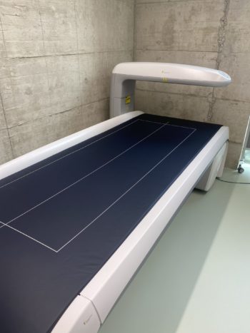
Bodyplethysmography (Lung function test)

Aim of the examination: A bodyplethysmograph is a device with great diagnostic value to detect limitations of the lungs on the one hand and to carry out follow-up examinations on the other hand. This method is therefore ideally suited for detecting possible long-term consequences after COVID-19.
Procedure of the examination: You will perform a series of lung function tests while wearing a nose clip and breathing in and out through a mouthpiece. During the tests, you will also be in the booth shown in the picture. The door is only closed during the first test (approx. 3 min). This is used to measure the breathing resistance at rest and the different lung volumes. After a sufficient break, the classic spirometry follows, in which the ability to exhale quickly and forcibly is tested. Finally, the diffusion capacity of the lungs is examined. This reflects how well the transfer of oxygen from the lungs into the blood works. In this test, you inhale a very small amount of carbon monoxide. This amount is completely safe for you.
Duration of the examination: Approximately 30 minutes.
Strength and balance tests

Aim of the examination: Numerous studies with post-COVID-19 patients report reduced muscle strength and muscle mass. This may be a consequence of physical inactivity but also of COVID-19 directly. Muscle strength and mass are important predictors of general mortality and central to healthy ageing. Reduced muscle strength and mass can also affect endurance performance (see cardiopulmonary exercise testing) and also quality of life. In the planned study, we want to find out to what extent muscle strength is affected by COVID-19. Patients with persistent symptoms also report cognitive problems. We would like to quantify one aspect of this problem by means of a balance test.
Procedure of the examination: The participants perform two strength tests and a balance test. In the first strength test, measuring hand grip strength, the aim is to grip an implement with the dominant hand as tightly and as quickly as possible. The second test measures leg strength. You exert as much force as possible with bent legs against a resistance that you hold with your hands. The balance test involves standing as still as possible for 10 seconds with your legs bent (in a tandem stance) on a force plate.
Duration of the examination: Approximately 15 minutes.
Body composition
Aim of the examination: Numerous studies with patients post-COVID-19 report reduced muscle mass up to one year after infection. Reduced muscle mass is not only important for the functioning of the body but also for overall mortality.
Accurate measurement of body composition can distinguish between fat and muscle mass and also provide information on bone density (conventional scales cannot). Therefore, it is very useful for documenting the current status but also for tracking progress.
Procedure of the examination: A determination of the body composition (muscle mass, fat mass and bone density) is made by means of Dual Energy-X-Ray-Absorptiometry, DXA for short. This device is a low-radiation X-ray method. The radiation exposure is much lower than with a normal X-ray and the examination only takes a few minutes. The measured resistances in the body can be used to estimate muscle mass, fat mass, protein and mineral content as well as water distribution. Even the fat mass in the abdominal cavity can be determined. In connection with diabetes, this represents an unfavourable metabolically active fat tissue and an important marker of “metabolic health”.
Duration of the examination: Approximately 15 minutes.
Vascular function (Micro- and macrocirculation)
Flow-mediated dilation (macrocirculation)
Aim of the examination: Via the measurement of flow-mediated vasodilation (FMD), the ability of the artery to dilate in response to increased blood flow is examined.
Vasodilation, the ability of the artery to dilate in response to increased blood flow is examined. The more pronounced the dilation is after congestion, the better the function of the artery wall. Dilatation depends on the ability of the inner layer cells (endothelial cells) of the brachial artery to produce nitric oxide (NO), which relaxes the vascular smooth muscle cells of the artery wall, thereby reducing tone in the vessel wall and subsequently allowing the artery to dilate more.

Procedure of the examination: The measurement of flow-mediated vasodilation (FMD) is carried out with harmless ultrasound on the brachial artery. The blood flow in the forearm is restricted for 5 minutes using a blood pressure cuff and subsequently released. The cuff is inflated above systolic blood pressure during the inflation phase, so that little blood can flow into the forearm. This is harmless to the arm and does not usually cause any pain. The diameter of the artery is measured before and after 5 minutes of congestion and for 3 minutes after the congestion is relieved. The measurement is made with self-adjusting ultrasound heads and automatic migration detection software, which together provide the most reproducible measurement results.
Duration of the examination: The examination lasts approx. 30 minutes, including 5-10 min preparation, 15 min resting phase in supine position before measurement and approx. 10 min measurement phase.
Retinal analysis (Microcirculation)

Aim of the examination: The examination of the fundus of the eye examines the diameters of the small arteries and veins of the retina, which are ~100-250 µm. The diameters of the retinal vessels and the ratio of arteries to veins (AV ratio) reflect the risk of cardiovascular diseases. The so-called cardiovascular risk increases in particular with narrow retinal arteries and wide veins. With this simple method, the health of the smallest vessels (microvascular) can be assessed representatively by looking into the eye. In addition, similar to the vascular examination of the arm, the dilatability of the retinal vessels can also be examined. This is done by means of flicker light stimulation. The examinations are used in prevention for screening the cardiovascular risk and for monitoring the progress of drug and non-drug therapies (physical activity and medical training therapy). In the COR-PHYS study, the examinations serve to identify possible impairments of the microcirculation by COVID-19.
Procedure of the examination: The retinal vascular analysis is an eye examination similar to what you know from the ophthalmologist. During the second part of the measurement, flicker light is used to provoke a vascular reaction. The images of the ocular fundus are stored and evaluated in a standardised way at a later time by an experienced examiner using special software.
Duration of the examination: Approximately one hour.
Echocardiography
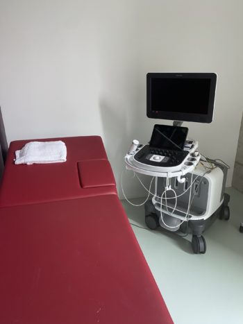
Aim of the examination: The ultrasound examination of the heart is also called echocardiography. It is used to non-invasively assess the structure and function of the heart as a central circulatory pump. From a sports medicine/internal medicine point of view, the systolic (ejection performance) and diastolic function (filling phase) are important. For this reason, ultrasound is used to examine the heart muscle as well as the heart valves more closely. The restriction of the diastolic function is caused, for example, by a stiffening of the heart muscle, as can be observed in long-term high blood pressure or also in heart weakness (cardiac insufficiency). Another important parameter for assessing the heart is the wall thickness. In athletes, the wall thickness of the left ventricle is in harmonic proportion to the internal diameter of the left ventricle. A relative increase is often seen in hypertension or in anabolic steroid abuse. A relative decrease in wall thickness is seen, for example, in heart failure, in which the ejection capacity of the heart is additionally limited in advanced severity. In sports medicine, cardiac structure and function are always assessed in connection with ergometric performance (maximum oxygen uptake, lactate performance diagnostics) and the exercise ECG.
Procedure of the examination: Echocardiography is performed while lying down using ultrasound, which is imperceptible and harmless through the chest wall. It is the same ultrasound that is used for pregnant women. The heart is examined from different standardised angles and Doppler methods (M-, B-mode, pulse, tissue Doppler). The transducer is placed on the skin and the contact is optimised by ultrasound gel. Suitable images and clips (mini films lasting a few seconds) are stored for evaluation. You can follow part of the examination on the screen.
Duration of the examination: Approximately 30 minutes.
Wearables
24 h blood pressure measurement
Aim of the examination: According to custo med, up to one-third of all hypertension patients remain undetected or receive limited therapy with conventional blood pressure measurement on the upper arm. In the long-term blood pressure measurement, only the central blood pressure provides a comprehensive picture of the actual blood pressure situation, as it directly affects the organs. In addition, the central pulse wave velocity can be recorded, which serves as an important marker for the stiffness of your arteries.
Procedure of the examination: At the same time as the vascular examination, a first measurement is taken under standardised conditions while you are lying down. At the end of the first study appointment, you will then be equipped with a 24 h blood pressure monitor and instructed on how to use it. You will wear this device for the next 24 hours.
Duration of the examination: 24 hours.
Accelerometry
Aim of the examination: The objective measurement of sporting and everyday movement with a movement sensor is indispensable today for a well-founded recording of physical activity. Only on this basis can reliable, individual statements about physical activity be made. Compared to a questionnaire, objective movement measurement is much more precise, because people usually estimate themselves to be much more active with a questionnaire than they really are. Furthermore, it can also be used to record the quality of sleep.
Procedure of the examination: The movement sensor is worn like a watch on the wrist for 24 hours a day over the entire measurement period (8 days). It is not a hindrance to everyday or office activities and is hardly noticeable to others. It is kept on for showering or swimming. The device does not need to be charged.
During the second study visit, the motion sensor is programmed, explained and given to the patient (10 minutes). The device is then worn for 8 days and all activities are recorded during this time. Afterwards, the motion sensor is returned with a pre-paid envelope. The recorded data will then be analysed by our team.
In addition, you will be asked to wear another fitness wristband on your other wrist. This records important bodily functions for us such as heart rate, heart rate variability, respiratory rate and oxygen saturation.
Questionnaires

Aim and procedure of the examination: The following information is collected in order to comprehensively document possible long-term effects as well as their severity and possible influencing factors for the above-mentioned measurement parameters. These influencing factors will later be taken into account in the statistical analyses.
Using digital questionnaires, the following data is collected:
- disease course,
- post-COVID-19 symptoms,
- physical activity,
- quality of life,
- fatigue,
- dyspnoea,
- depression stress, and anxiety
- post-exertional malaise
- postural orthostatic tachycardia syndrome.
Duration of the examination: Two times approximately 10 minutes.
Further details on the study design and the tests performed will be published in the study protocol.